1.2 Tesla High field open MRI

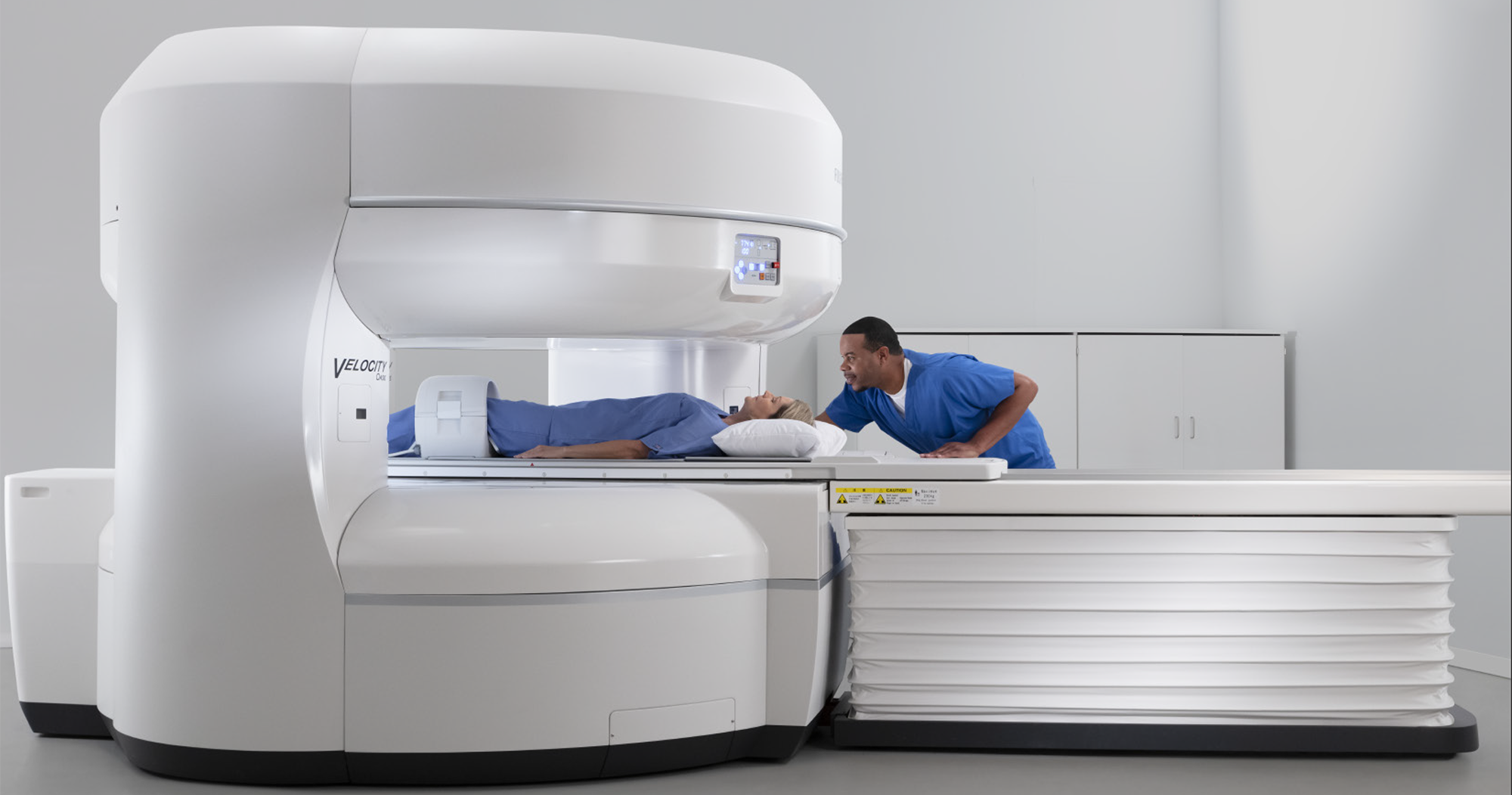
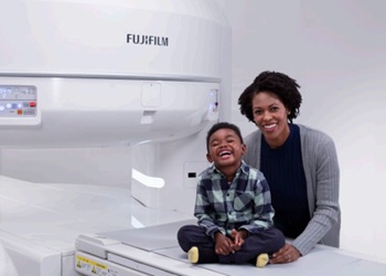
Introducing the 1.2 Tesla High Field Open MRI System: Advancing Accessibility and Comfort in Imaging.

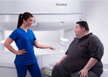
The 1.2 Tesla High Field Open MRI System heralds a new era in medical imaging, offering a combination of high magnetic field strength and an open design for enhanced patient comfort and accessibility. Unlike traditional closed-bore MRI systems, the open design of the 1.2 Tesla High Field MRI System provides patients with a more spacious and less restrictive scanning environment, reducing feelings of claustrophobia and anxiety during the imaging procedure.

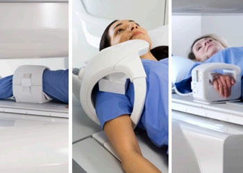
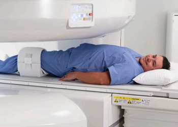
At its core, the 1.2 Tesla High Field Open MRI System utilizes a magnetic field strength of 1.2 Tesla to generate detailed and precise images of the human anatomy. This high field strength, coupled with advanced imaging technology, allows healthcare providers to obtain clear and accurate images of soft tissues, organs, and structures, aiding in the diagnosis and treatment of a wide range of medical conditions.

The open design of the MRI system features a larger and more accommodating bore, or tunnel-like opening, which can accommodate patients of various body types and sizes. This inclusivity ensures that all patients, including those who may have difficulty undergoing imaging in a traditional closed-bore MRI, can receive the diagnostic information they need with comfort and ease.

In addition to its patient-friendly design, the 1.2 Tesla High Field Open MRI System offers versatility across a spectrum of clinical applications. From neuroimaging and orthopedics to abdominal imaging and oncology, this advanced system can perform a wide array of imaging studies, providing valuable insights for healthcare providers and patients alike.

Furthermore, the open design of the MRI system enhances workflow efficiency and patient throughput in healthcare facilities. Technologists have improved access to patients during the scan, enabling them to monitor patient comfort and positioning more effectively. This streamlined approach not only reduces scan times but also enhances overall patient satisfaction and experience.

In summary, the 1.2 Tesla High Field Open MRI System represents a significant advancement in medical imaging technology, bridging the gap between high field strength and patient comfort. With its open design, versatile imaging capabilities, and emphasis on accessibility, this innovative MRI system is poised to transform the landscape of diagnostic imaging, improving outcomes and experiences for patients and healthcare providers alike.

Patient Comfort

The Velocity Open MRI offers a comfortable experience for even the most diverse patient
population.

Velocity also provides:

  • 1.2 Tesla strength
  • 270º viewing angle for the best scanning experience
  • Patient lighting that enhances the openness of the machine passageway
  • Noise reduction without compromising scanning capability
  • Customizable audio that can use the patient’s own music
  • A wide patient table for comfortable, easy positioning
  • A lower table for easy access by wheelchair patients
  • Patient accommodation up to 660 lbs
Orthopedic Imaging

The Velocity Open MRI delivers comfortable extremity imaging. Wrists and elbows are imaged at the patient’s side. Comfortable
isocenter shoulder imaging is easily managed for all patients.

Velocity also provides:

  • Unlimited lateral opening to accommodate the broadest patients
  • Excellent fat suppression even with comfortable positioning
  • Easy ABER (Abduction External Rotation) positioning with the Shoulder coil for optimal labrum visualization
  • Lateral movement to simplify positioning
  • Streamlined flexion/extension imaging that differentiates your capabilities
  • HOAST and regional shimming for superb RF fat saturation
  • Techniques for imaging around prosthesis
Orthopedic Imaging

The Velocity Open MRI delivers comfortable extremity imaging. The 1.2 Tesla MRI magnet
provides quality of image not previously available in an Open MRI.

Velocity also provides:

  • Unlimited lateral opening to accommodate the broadest patients with easier positioning, easier tolerance
  • Wrists and elbows imaged at the patient’s side
  • Easy ABER (Abduction External Rotation) positioning with the Shoulder coil for optimal labrum visualization
  • Lateral table movement for easier isocenter positioning on all extremity exams
  • Excellent RF fat saturation
  • Techniques for imaging around prosthesis
Vascular Imaging
Vascular imaging on the Velocity Open MRI provides quality scans without compromising patient comfort. Capabilities include:
  • 2D and 3D TOF for intra-cranial and extra-cranial imaging
  • FLUTE™ ensures arterial phase capture
  • TPEAKS™ centric k-space filling
  • TRAQ MRA depicts blood flow dynamics
  • PVA with multi-coil connections, auto table movement, and large FOV
  • VASC™ non-contrast MRA for renal, carotid and peripheral applications
  • Dedicated RF Coils provide outstanding coverage and uniformity
  • Volume Rendering and auto MIP preview
Neurological Imaging

Neurological imaging on the Velocity Open MRI puts both patients and technologists at ease. The open design reduces claustrophobia.

In addition:

  • Multi-plane imaging can be accomplished without repositioning the patient and
    without volume CT’s ionizing radiation.
  • Circle of Willis can be obtained by Velocity’s ultra fast scanning capabilities in one acquisition.
  • Diffusion Weighted Imaging can detect an infarct in the sub acute stage, as well as distinguish between acute and chronic lesions.
  • RADAR radial acquisition reduces patient motion artifact, yielding diagnostic quality scans in even the most difficult to image patients.
Bariatric Imaging

More than 64% of U.S. adults are overweight or obese. This number is increasing steadily.
Velocity High-field MRI meets the bariatric imaging challenge, delivering:

  • Industry-best table capacity of 660 lb to accomodate diverse patients
  • Wide patient table of 32” that places anatomy comfortably at iso-center for optimal SNR
  • 3-axis motorized lateral table movement which provides convenience
  • Table that lowers to 20” for easy access 
  • Sensitive multichannel RF coil technology to adjust to all patients
Pediatric Imaging

Constant parent-child contact differentiates your pediatric MRI service with Velocity. Even the smallest patients have the ideal MRI experience.

In addition:

  • Open architecture allows pediatric patient security and comfort, sometimes eliminating
    the need for sedation or anesthesia
  • Halo coil delivers quality imaging and an all-around view
  • TPEAKS™ centric k-space filling
  • Unlimited patient access increases efficiency
  • Parents can talk to and reassure the child during exam
  • RADAR motion-free imaging eliminates repeat scans and improves image quality
  • RADAR radial acquisition reduces patient motion artifact, yielding diagnostic quality scans in even the most difficult to image patients.
  • Fast scanning techniques keep study time minimal
  • SoftSound gradient technology reduces acoustic noise
Geriatric Imaging

The open design of the Velocity Open MRI maximizes comfort and ease for geriatric patients. Patients can have a friend or loved one with them throughout the exam.

In addition:

    • Technologist has access to the patient from
      anywhere around the magnet
    • Technologist can easily observe the patient
      during the exam
    • Novel blanket coils allow for quick positioning
    • In-bore lateral movement simplifies iso-center
      positioning for all extremities
    • Motion-compensated RADAR translates into
      excellent diagnostic sequences even with
      difficult patients
    • SoftSound gradient technology reduces
      acoustic noise
Abdominal Imaging

Unparalleled patient comfort, exceptional abdominal image quality and ultra-fast scan times are now available in an open environment with the Velocity 1.2T High-Field Open MRI system.

In addition:

  • TIGRE™ for T1-weighted 3D fat-suppressed gradient echo dynamic imaging
  • 2D and 3D MRCP applications
  • TPEAKS™ centric k-space filling
  • Diffusion-Weighted Imaging applications
  • RADAR™ motion compensation for free breathing applications
  • RAPID™ Parallel Imaging
  • VASC-ASL™ for non-contrast MRA applications
  • FLUTE™ Fluoro-Trigged MRA and TRAQ™ Time-Resolved MRA for contrast-enhanced
    vascular studies