MRI Associates became established in early 1995 and began by offering MRI and X-Ray. After recognizing the needs of our patients and developing relationships within our Medical Community, we expanded our services. We have expanded into 8 cities: Palm Harbor, Lakeland, Winter Haven, Brandon, Sarasota, Venice, Bradenton, and Port Charlotte. Palm Harbor and Highland offer 2 high field systems, with one being a Wide Bore MRI for claustrophobic pateints. In Sarastoa we have a 1.2 High Field Open MRI that is one of the only available high field open MRI systems in Florida. It is an amazing option for truly claustrophobic patients.
Magnetic resonance imaging (MRI), ultrasound, CT, x-ray and other diagnostic testing plays a key role in identifying and managing health conditions, diseases and illnesses. At MRI Associates, we use the latest techniques and technology to provide healthcare practitioners and patients with the data they need to make informed care decisions. As a premier provider of diagnostic services in and around Tampa Bay, we pride ourselves on our attention to every patient we serve, offering complete, compassionate care to ensure patients feel comfortable and confident throughout their experience with us.
We also make it easy for physicians to get the critical information they need by implementing a user-friendly environment for accessing MRI and other diagnostic results and patient information while implementing secure, HIPPA-compliant practices that ensure the highest degree of confidentiality and privacy.
“The Patient is our number one priority. We pledge to provide a professional environment with exceptional technology where you will always receive the highest quality care. At MRI Associates, we understand that dealing with a health issue can be a nerve-wracking experience for patients, and we strive to ensure the services and environment we offer are 100% patient-centered.”
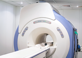
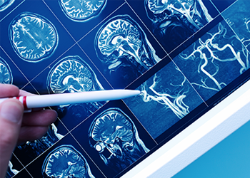
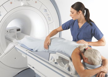
Magnetic Resonance Imaging (MRI) is a method of obtaining detailed pictures of internal body structures without the use of radiation or radioactive substances of any kind. This is accomplished by placing the patient in a magnetic field while harmless radio waves are switched on and off. All of our locations have High Field 1.5 Tesla Magnet Strength Systems.
You can help to produce a high quality image by lying still during the examination while breathing normally. The average scan takes 5 to 15 minutes – the complete examination about 30 to 45 minutes – during which several dozen images will be produced.
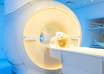
Our NEW Wide OPEN Short Bore 1.5 T High Field MRI System allows us to offer our patients the optimized comfort of conventional open bore systems (Open MRI), as well as the high-quality imaging of conventional closed bore systems (High Field MRI). Wide bore MRIs have broadened the patient demographics of those who can be tested (Weight capacity: 550 lbs). The wider bore makes the exam a good option for claustrophobic, broad shouldered, or weight challenged patients.
Most scans allow the patient’s head to be out of the scanner to help with those who may be claustrophobic.
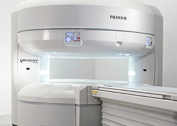
Open MRI is used to accommodate claustrophobic, obese and pediatric patients. Conventional MRI scanners are a cylinder shape, while an open MRI does not completely surround your body. It is open on two of its sides. An Open MRI provides a more relaxed, less confining environment and lower noise levels making it less stressful for the patient. MRI Assocaites has a True High Field Open MRI Systems at Sarasota MRI.
The Oasis Velocity supports up to 660 lb patients on an 83 cm wide table. A true open design, patients have a massive 270° unobstructed view for maximum comfort.
MRI Arthrogram studies are used to obtain detailed images after a contrast material has been injected into the affected joint. The resulting image shows the soft tissue structures of the joint to find the cause of ongoing, unexplained joint pain, swelling or abnormal movement. Arthrography is done most commonly to identify abnormalities associated with the shoulder, wrist, hip, knee and ankle. Patients who undergo this procedure usually have complained of persistent, unexplained joint pain or discomfort. Arthrography images may allow identification of problems with a joint’s function or indicate a need for a joint replacement.
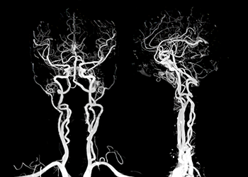
MRI Angiography (MRA) is an MRI study of the blood vessels. It utilizes MRI technology to detect, diagnose and aid the treatment of heart disorders, stroke, and blood vessel diseases. MRA provides detailed images of blood vessels without using any contrast material.
Magnetic resonance venography (MRV) is the most sensitive and specific test for the assessment of deep and superficial venous disease in the lower legs and pelvis, areas not accessible by means of other modalities. MRV is particularly useful because it can help detect previously unsuspected nonvascular causes of leg pain and edema when the clinical presentation erroneously suggests venous insufficiency or venous obstruction.
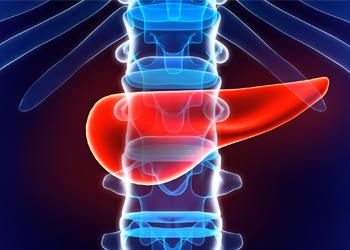
MRCP test is a specialized MRI exam that evaluates the hepatiobiliary and pancreatic systems, including the liver, gallbladder, bile ducts, pancreas and pancreatic duct. MRCP stands for Magnetic Resonance Cholangiopancreatography:
Cholangio = bile vessel
Pancreato = pancreas
Graphy = image
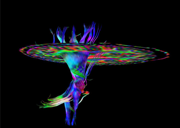
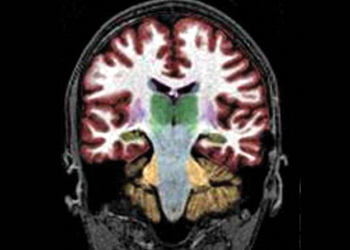
Diffusion tensor imaging (DTI) can detect abnormalities that significantly relate to long-term outcomes in mild traumatic brain injury (TBI).
DTI is an innovative diagnostic technique is a variation on the standard MRI that is used to create images of internal organs through magnetic images. The DTI isolates water movement within the brain, which allows doctors to isolate regions that are not functioning properly. Traditional MRI scans cannot highlight these abnormalities, because they do not have the capability of tracking water molecules in the same way.
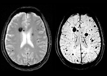
Susceptibility Weighted Imaging (SWI) is a cutting-edge technology that uses extremely high-resolution imaging to detect damage to the brain caused by microhemorrhages, shearing and diffuse axonal injuries. The technology was originally developed to map the brain’s venous architecture. This high-resolution imaging allows medical professionals to see even the smallest lesions in the brain. By exploiting the susceptibility differences between tissues, SWI’s high-resolution enhanced-contrast imaging maps areas of the brain that exhibit venous blood, hemorrhage and iron storage.
NeuroQuant® can be a very effective tool to show traumatic brain injury resulting from carbon monoxide exposure. Carbon monoxide exposure causes diffuse brain injury, meaning multiple lobes throughout the brain lose volume (injury) as oppopposed to a focal injury in a specific location, such as when the front and back portions of the brain are injured during a whiplash-type mechanism of injury. Measured loss of volume in the lobes diffusely throughout the brain can objectively prove that a person suffered a brain injury as a result of carbon monoxide exposure.
Breast Magnetic Resonance Imaging (MRI) is another non-invasive breast imaging tool that produces detailed images of the breast. Breast MRI utilizes a magnetic field and radio waves as well as an intravenous injection of a contrast agent to highlight breast abnormalities.
Breast MRI is intended to be used in addition to a mammogram or another breast-imaging test, and not as a replacement for a mammogram. Although it is highly sensitive, breast MRI can still miss some breast cancers that a mammogram can detect.
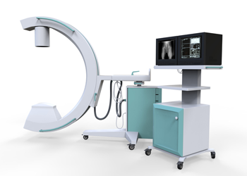
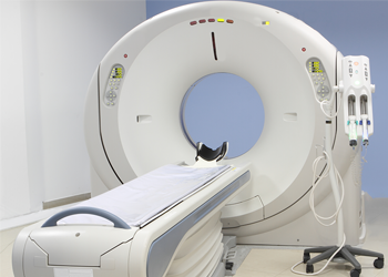
CT scan; Computed Axial Tomography (CAT) is a method of body imaging in which a thin x-ray beam rotates around the patient. Small detectors measure the amount of x-rays that make it through the patient or particular area of interest.The CT scanner looks like a large doughnut with a table that extends into the opening of the machine. CTs are used for a variety of tests that can image every part of your body. These scans can help in the diagnosis of infections, cancer, heart disease, fractures and many other conditions. MRI Associates offers patients modern technology with CT scanner in each of our seven facilities.
Computed Tomography Angiography (CTA) uses an injection of contrast material into your blood vessels and CT scanning to help diagnose and evaluate blood vessel disease or related conditions, such as aneurysms or blockages. When a contrast material is introduced to the bloodstream during the procedure, it clearly defines the blood vessels being examined by making them appear bright white. Some reasons to have a CTA include:
• To find an aneurysm or forms of plaque in the walls of arteries
• Abnormal blood vessel formations inside your brain
• Identify blood vessels damaged by injury, tumors or find blood clots
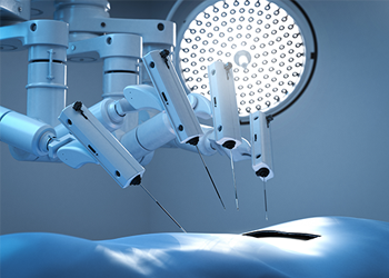
3D CT Based Planning begins with a CT scan of an extremity. Those detailed images taken from multiple anges are then used to create 3D images of the shoulder, hip, knee, ankle, etc. This allows for the Smart Robotics to show the surgeons precise cut points for insured accuracy. These 3D models are created specifically for each and every patient, each 3D model is custom to the patient and the procedure the surgeon is performing. This allows the surgeon to make sure the pre-operative planning is perfect for the patient and their unique anatomy.
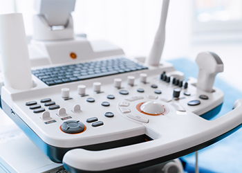
Ultrasound imaging is a common diagnostic medical procedure that uses high-frequency sound waves to produce dynamic images (sonograms) of organs, tissues, or blood flow inside the body. The test is done in our Ultrasound room. You will be lying down for the procedure. A clear, water-based conducting gel is applied to the skin over the area being examined to help with the transmission of the sound waves. A handheld probe called a transducer is then moved over the area being examined. You may be asked to change position so that other areas can be examined.
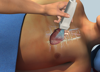
Echocardiogram, often referred to in the medical community as a cardiac ECHO or simply an ECHO, is a sonogram of the heart. Also known as a cardiac ultrasound, it uses standard ultrasound techniques to image two-dimensional slices of the heart. The latest ultrasound systems now employ 3D real-time imaging.
In addition to creating two-dimensional pictures of the cardiovascular system, an echocardiogram can also produce accurate assessment of the velocity of blood and cardiac tissue at any arbitrary point using pulsed or continuous wave Doppler ultrasound. This allows assessment of cardiac valve areas and function, any abnormal communications between the left and right side of the heart, any leaking of blood through the valves (valvular regurgitation), and calculation of the cardiac output as well as the ejection fraction.
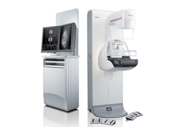
Mammograms are specialized X-ray examinations of the breast. Two types of mammogram studies are commonly performed. A SCREENING MAMMOGRAM is performed on women who have no current symptoms or breast problems while a DIAGNOSTIC MAMMOGRAM is performed specifically to evaluate a breast problem or revisit a previous abnormal finding.
MRI Associates offers 3D Mammography at Palm Harbor MRI, Winter Haven MRI and Brandon MRI all have State- of-the-Art Fuji 3D Mammogram systems.
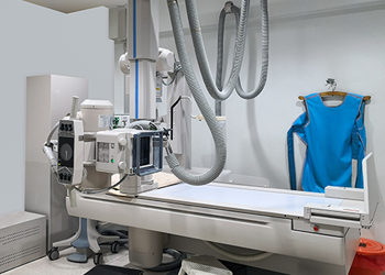
An X-ray examination uses electromagnetic radiation to make images of your bones, teeth and internal organs. Simply put, an X-ray allows your doctor to take pictures of the inside of your body.One of the oldest forms of medical imaging, X-ray is a painless medical test that can help your doctor in diagnosis and treatment.
X-ray exams also play an important role in the detection and diagnosis of cancer. In fact, one use of X-ray in diagnosing cancer is to see whether you have lung cancer or whether cancer from another part of the body has spread (metastasized) to the lungs. Cancer may appear lighter in color on X-ray films than does normal, healthy lung tissue. X-rays may also be used to examine cancers of the intestines, stomach, liver, spleen, kidneys and breasts.
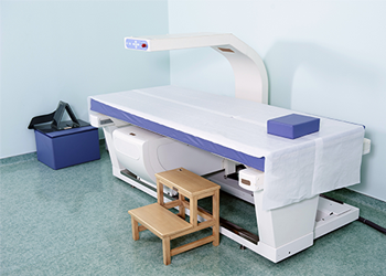
Bone Densitometry (DEXA) DEXA (short for dual-energy x-ray absorptiometry) is an advanced technology that safely, accurately and painlessly measures bone mineral density. It helps determine whether you are at high, increased or low risk of fracturing a bone.
DEXA is often performed on the lower spine and hips. In children and some adults, the whole body is sometimes scanned. Peripheral devices that use x-ray or ultrasound are sometimes used to screen for low bone mass, mostly at the forearm.
Diagnostic Health Screenings can help identify health risks prior to symptoms. Our imaging services take a peek at vital sections of the body and interprets if there are any risks associated with the images.
Early detection & knowledge is a powerful tool for the health and well being of all our patients.
A Board Certified Radiologist will review all images and provide a Radiology Report to your Referring Physician at no extra cost. All studies are completed without contrast.
To learn more about our services, explore our site or call us at one of our seven convenient locations today.
Main Office:
PALM HARBOR MRI
32615 US Hwy 19 North
Palm Harbor, FL 34684
Phone: 727.787.6900
Fax: 727.216.4789