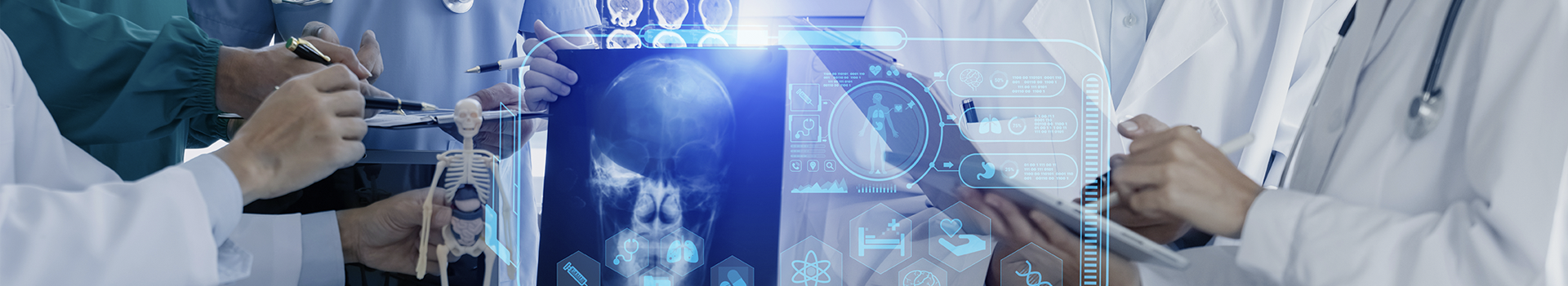
CT scan; Computed Axial Tomography (CAT) is a method of body imaging in which a thin x-ray beam rotates around the patient. In CT Scan process small detectors measure the amount of x-rays that make it through the patient or particular area of interest.
A computer analyzes the data to construct a cross-sectional image. These images can be stored, viewed on a monitor, or printed on film, or disk. In addition, three-dimensional models of organs can be created by stacking the individual images, or “slices.”
Why the test is performed:
CT provides rapid, detailed cross-sectional imaging of the patient which can then be reconstructed into three-dimensional models, as needed. Intravenous contrast enhanced scans allow for evaluation of vascular structures and further evaluation of masses and tumors.
CT is often utilized in the trauma setting to evaluate the brain, chest, and abdomen. As well, CT can be used to guide interventional procedures, such as biopsies and placement of drainage tubes.
CT scan process:
You will be asked to lie on a narrow table that slides into the center of the scanner. Depending on the study being performed, you may need to lie on your stomach, back, or side. If contrast dye is to be administered, an IV will be placed in a small vein of a hand or arm.
Much like standard photographic cameras, any motion you make causes blurred images in CT. Therefore, the operator will give you instructions through an intercom on when to hold your breath.
As the exam takes place, the table will advance small intervals through the scanner. Modern “spiral” scanners can perform the exam in one continuous motion. Generally, complete scans will only take a few minutes. However, additional contrast-enhanced or higher-resolution scans will add to the scan time. The newest multi detector scanners can image your entire body, head to toe, in less than 30 seconds.
Procedure of CT scan:
You may be asked to drink contrast immediately prior to the CT scan, or 4 to 6 hours beforehand. The contrast may be non-reactive, chalky-tasting barium sulfate, which will eventually pass in the stools, or absorbable clear Gastrografin solution. You may also be asked to fast (no solids or liquids) for 4 to 6 hours if contrast dye is to be used. Our contrast is mixed with apple juice.
Since x-rays have difficulty passing through metal, the patient will be asked to remove jewelry and wear a hospital gown during the study.